Dear all,
Some interesting reviews and research this month. Will separate blog from now into clinical studies (i.e. treatments and risk factors) and other interesting stuff.
Hope you enjoy
All the best
Peter
Studies to apply to your practice
Mani-Babu et al. have performed an excellent systematic review of the effectiveness of shockwave therapy in lower limb tendinopathy. There is some evidence shockwave is superior to home exercise and steroid in gluteal tendinopathy & patellar tendinopathy. The issue is many of these patients have already failed exercise, and the quality of the exercise needs to be questioned. There is evidence that shockwave is superior to eccentric for insertional Achilles, which make sense given through range isotonic loading is usually not good for these. There is evidence that adding exercise to shockwave is superior to shockwave alone in midportion Achilles. I am a big believer in this approach, ie shockwave as a pain modulator accompanied by GOOD rehab. Some studies show shockwave may be no better than placebo (it’s a very sexy and impressive machine, not many people have it, and it makes loud noises = maximum placebo), which reinforces the importance of using it as an adjunct to some form of loading.
http://ajs.sagepub.com/content/early/2014/05/08/0363546514531911.abstract
Dingemanse et al. have reviewed two previous reviews and twenty RCT’s investigating the effect of electrophysical modalities in treating lateral elbow tendinopathy. 70% of the RCT’s were reported to be high quality, which is impressive, but high quality was >50% of their quality scale. There was no evidence or conflicting evidence for shockwave cf placebo, pulsed electromagnetic field vs placebo, laser vs placebo, US vs no treatment. On the other hand there was limited evidence for exercise cf US and short-term effects of noxious inputs on trigger points, ie electrical stimulation. Overall, the evidence for passive treatments on medium to long-term outcome is very limited at best, but interventions that can reduce pain in the short term should be explored further.
http://www.ncbi.nlm.nih.gov/pubmed/23335238
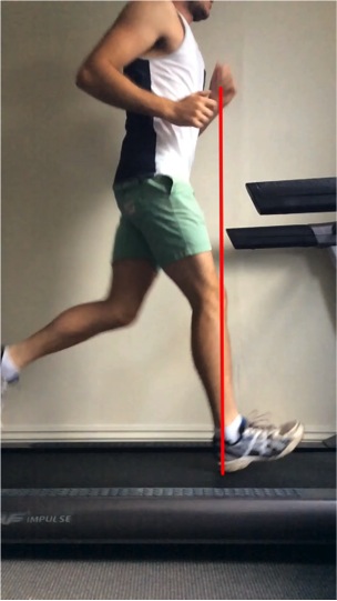
Lorimer et al. have reviewed running related risk factors for Achilles tendinopathy. Risk factors that were clearly associated included softer surfaces like sand and track, training near race pace (so faster), greater arch (protective effect), greater breaking force (perhaps associated with overstride). Excessive or prolonged pronation was NOT clearly linked with Achilles injury, but faster rate of pronation was from one study. I suspect running mechanics (e.g. overstride, heel strike) is associated with breaking force and rate of pronation, suggesting running re-training has a significant role in Achilles management.
http://www.ncbi.nlm.nih.gov/pubmed/24898814
Interesting narrative review by Alfredson. I agree with his strong reservations regarding stem cells, PRP into tendons. Also agree with thoughts on targeting tissue in the ventral Achilles with surgery, rather than the usually intratendinous surgery that often leads to huge tendon thickening and has a long recovery time. His overall message seems to be leave the tendon alone, and if you want to/need to interfere (ie add something other than rehab), focus on the surrounding/ventral tissue.
https://www.oapublishinglondon.com/article/1117
Other interesting studies
Interesting study by Ngomo et al. showing decreased excitability of the motor cortex on the affected side among patients diagnosed with rotator cuff tendinopathy. The asymmetry in excitability between sides was associated with pain chronicity (>12 months). Supports recent findings by Rio et al. (2013 abstract) showing greater cortical inhibition in patellar tendinopathy. Suggests cortical neuroplasticity with injury, so we need to think broadly regarding mechanisms of dysfunction and focus treatments appropriately.
http://linkinghub.elsevier.com/retrieve/pii/S1388245714003162?via=sd
Rio E, Kidgell D, Moseley L, et al: Exercise to reduce tendon pain: A comparison of isometric and isotonic muscle contractions and effects on pain, cortical inhibition, and muscle strength. Journal of Science and Medicine in Sport. 2013, e28
Masood et al. investigated calf EMG and strength following 12 weeks of eccentric training in 10 Achilles patients and 10 controls. Muscle force and glucose uptake increased in both groups (force production was lower in painful group at baseline). Achilles glucose uptake was higher in Achilles pain group, but did not change with rehab in either group. Soleus EMG was higher in the painful group, and lateral gastroc EMG increased in this group after training. Findings suggest higher metabolism in Achilles tendinopathy, and higher soleus EMG probably due to reduced force potential.
www.ncbi.nlm.nih.gov/pubmed/24855138
Pearson et al. have reviewed of region specific tendon properties. Patellar tendon pathology is generally located around the proximal posterior tendon. Based on the moment arm of the patellar tendon, the proximal posterior tendon should be stress shielded (ie under less stress than anterior fibres. There is debate about this. Basso et al 2002 found strain of the posterior fibres was greater in an in vitro cadaver knee. Dillan et al. 2008, found greater posterior fibre load using an optic fibre technique. Almekinders et al 2002, found the opposite, greater anterior fibre load and stress shielding of the posterior fibres! They also used a cadaver model but less knee flexion (60 vs 90 degrees). Isolated tendon fascicles from the posterior tendon have been shown to be less stiffer (weaker) (Hansen 2008, Haraldsson 2005). If we assume there is stress shielding, what can we do about it clinically? Loading into greater ranges knee flexion ranges is an issue due to pain. So heavy heavy mid range loading best to adapt, even though anterior fibres may be under greater load.
www.ncbi.nlm.nih.gov/pubmed/24838651
Almekinders LC, Vellema JH, Weinhold PS. Strain patterns in the patellar tendon and the implications for patellar tendinopathy. Knee Surg Sports Traumatol Arthrosc. 2002;10(1):2–5.
Basso O, Amis AA, Race A, et al. Patellar tendon fiber strains: their differential responses to quadriceps tension. Clin Orthop Relat Res. 2002;400:246–53.
Dillon EM, Erasmus PJ, Mu ?ller JH, et al. Differential forces within the proximal patellar tendon as an explanation for the characteristic lesion of patellar tendinopathy: an in vivo descriptive experimental study. Am J Sports Med. 2008;36(11):2119–27.
Hansen P, Haraldsson BT, Aagaard P, et al. Lower strength of the human posterior patellar tendon seems unrelated to mature col- lagen cross-linking and fibril morphology. J Appl Physiol. 2010;108(1):47–52.
Haraldsson BT, Aagaard P, Krogsgaard M, et al. Region-specific mechanical properties of the human patellar tendon. J Appl Physiol. 2005;98(3):1006–12.
Bohm et al. et al. have shown that the Achilles tendon is stiffer (14%) and longer (distal soleus to calcaneum) (5%) on the dominant leg. They excluded people performing assymetric (e.g. fencing, high jump) and high performance sport. This finding may be explained by subtle favouring of the dominant leg over years. I expect there would be the opposite effect in jumping athletes who jump from their non-dominant side, as shown by Couppe et al 2008.
www.ncbi.nlm.nih.gov/pubmed/24798645
Couppé C, Kongsgaard M, Aagaard P, et al.: Habitual loading results in tendon hypertrophy and increased stiffness of the human patellar tendon. J Appl Physiol. 2008, 105:805-810.