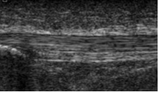
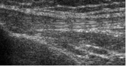
Tendon injuries affect many people and include tennis elbow, plantar fascitis, Achilles, patellar, tibialis posterior, as well as rotator cuff injury. Recent studies by our research group have shown that there are different phases of tendon injury on ultrasound imaging. Less severe pathology involves diffuse tendon (middle image) thickening, compared to the parallel and fibrillar appearance of normal tendon (left image). Severe pathology is characterised by a focal area of tendon degeneration (right image). Please read more about the study by clicking below.
The most important question is, does this influence how we treat these injuries? The simple answer is that it may. Diffuse thickening is probably representative of reactive tendinopathy symptoms and focal changes represent degenerative symptoms. These injuries are treated very differently so it is important to detect them
There is a big BUT. That is that pathology may be asymptomatic and severe focal degenerative pathology may present with acute, severe symptoms that are associated with reactive tendinopathy (i.e. acute on chronic). So, the most important consideration is the patients symptoms, followed by the imaging.


